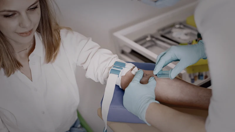
Yes. Stress can change blood test results. A short burst of fear can move cortisol, prolactin, white blood cells, plasma volume, glucose, lipids, and inflammatory signaling. Chronic stress can also change bone-marrow output and the way immune cells respond to cortisol. The key is timing: some values move within minutes, some over hours, and some only after months or years of repeated alarm.
Key takeaways
- A blood test is a snapshot of a living system, not a verdict carved in stone.
- The stress of waiting, the needle, or the result itself can alter the sample being collected.
- Cortisol and prolactin are especially sensitive to collection conditions and time of day.
- Acute stress can redistribute white blood cells and temporarily concentrate hemoglobin, hematocrit, protein, and lipids by reducing plasma volume.
- Glucose may rise or fall more slowly after a meal; the effect is stronger and more clinically relevant in diabetes.
- IL-6 can rise within hours; CRP is slower and is best read as a trend under comparable conditions.
- “Stress” should explain a mechanism—not erase a persistent abnormality.
You sit in the chair. The tourniquet tightens. The needle enters. You are trying to look calm, but the body has already made its decision.
Adrenaline rises. Blood vessels change tone. The heart pumps faster. Immune cells move. Cortisol begins its climb. Plasma shifts out of the circulation. The sample enters the tube carrying not only the chemistry of the previous month, but also the physiology of the previous five minutes.
That is why two samples from the same person can differ without either laboratory being wrong. One was taken after sleep, quiet, fasting, and twenty minutes in a chair. The other was taken after traffic, an argument, three hours of sleep, a sprint through the parking lot, and fear of what the number might say.
The useful question is not simply, “Can stress change bloodwork?” It can. The useful question is: which marker, in which direction, over what timescale, and by what mechanism?
First separate acute stress from chronic stress
Acute stress is the burst: the needle, the speech, the frightening message, the near miss, the argument outside the laboratory. It changes circulation and hormones within seconds to minutes.
Chronic stress is the alarm that keeps returning: months of caregiving, financial danger, hostility at home, fear at work, unresolved threat that has become the background of the day. It changes immune regulation, inflammatory output, sleep, metabolism, and the tissues that respond to cortisol.
These are not two unrelated processes. The chronic state is built out of repeated acute states plus incomplete recovery. The same alarm keeps sending the same instructions until the body begins to reorganize around them.
Which blood tests can move?
| Marker | What stress can do | Why | Typical window |
|---|---|---|---|
| Cortisol / ACTH | Often rises; chronic patterns may become high, flattened, or blunted. | HPA-axis activation; time of day and prior sleep strongly shape the result. | Minutes to hours |
| Prolactin | Can rise mildly during venipuncture or anticipatory distress. | Stress-related pituitary release; a rested sample may normalize. | Minutes |
| White blood cells | Total count and subtypes can shift rapidly; chronic stress can raise monocytes and neutrophils. | Adrenaline redistributes cells; sympathetic nerves can stimulate bone marrow. | Minutes to months |
| Glucose | May rise or remain elevated longer after a meal. | Adrenaline and cortisol mobilize fuel and reduce insulin effectiveness. | Minutes to hours |
| Hemoglobin / hematocrit / protein | Can appear temporarily higher. | Plasma volume falls, concentrating what remains in the tube. | Minutes |
| Cholesterol / triglycerides | Can appear higher during an acute stress task. | Much of the short-term rise can be hemoconcentration rather than new lipid production. | Minutes to an hour |
| IL-6 | Can rise after acute stress and climb faster under chronic stress. | Sympathetic and immune signaling activate inflammatory pathways. | Hours to years |
| CRP / hs-CRP | May run higher when chronic inflammatory signaling is higher. | The liver makes CRP downstream of IL-6; it moves more slowly than the immediate alarm. | Hours to days; trends matter |
Cortisol: the number most people expect to move
Cortisol is designed to move. It follows a daily rhythm, usually highest after waking and lower later in the day. Then an acute threat can add another rise on top of that rhythm.
In laboratory stress studies, public speaking and mental arithmetic reliably activate the hypothalamic-pituitary-adrenal axis and increase cortisol. But the collection procedure can become part of the experiment. Venipuncture is itself a potent stressor; people who experience a vasovagal reaction can begin with an abnormally high baseline and then appear not to respond to the later task because the needle already fired the system.[11]
Translation: a cortisol result is inseparable from clock time, sleep, food, medication, the collection method, and what happened immediately before the draw. A single number can be real and still represent the moment rather than the whole month.
Chronic fear does not always mean chronically high cortisol. Repeated activation can produce a flatter rhythm or a blunted response. The more important change may be in the receiver: immune cells can become less sensitive to cortisol's signal.
Sheldon Cohen and colleagues exposed healthy volunteers to rhinovirus after measuring long-term threatening stress and glucocorticoid resistance. Long-term stress was associated with reduced immune-cell sensitivity to cortisol; that resistance predicted more inflammatory cytokine production and greater risk of developing a clinical cold.[2]
The brake is still being pressed. The tissue has stopped listening.
Prolactin: the blood draw can manufacture the problem it is testing
Prolactin is one of the clearest examples of collection stress altering interpretation. A mildly high result can lead to repeat testing, medication review, pituitary imaging, and fear of a tumor. Yet the needle and the anticipation around it can raise the hormone.
In a hospital series of 93 people referred with elevated prolactin, levels fell during rested serial sampling in 78%. The final result returned to the normal range in 54%, consistent with stress-related hyperprolactinemia rather than persistent pathological elevation.[10]
This does not make every high prolactin “just stress.” It shows something more useful: the measurement protocol can separate a transient alarm from a persistent signal. When the first elevation is mild, the next step is often a cleaner measurement rather than an immediate biography of disease.
White blood cells: stress can move the army before infection arrives
A complete blood count looks objective because it counts cells. The count is objective. The location of the cells is dynamic.
During ten minutes of mental arithmetic, healthy women showed rapid increases in total leukocytes, lymphocytes, CD8 cells, and natural-killer cells.[4] Other human studies found leukocytosis across lymphocyte, monocyte, and granulocyte populations during speaking and exercise tasks.[5]
Adrenaline changes adhesion and traffic. Cells that were resting along vessel walls or in reservoirs enter the circulating sample. The tube therefore sees a different immune army even though no infection appeared in those ten minutes.
Chronic stress can go deeper. In intensive-care staff, Heidt and colleagues found higher circulating monocytes and neutrophils during stressful duty periods. Their animal work traced the mechanism into bone marrow: sympathetic noradrenaline changed the stem-cell niche and accelerated production of inflammatory leukocytes.[1]
That is why “stress leukocytosis” is biologically real—and why a persistent or marked elevation should still be followed until the actual cause is clear.
Hemoglobin, hematocrit, total protein, and lipids: sometimes the tube is more concentrated
Acute stress changes blood volume. Pressure rises inside the circulation, fluid shifts out of plasma, and the substances left behind become more concentrated. The result can imitate a sudden rise in red cells, hemoglobin, hematocrit, total protein, cholesterol, and triglycerides.
In a randomized experiment, twenty minutes of frustrating mental work increased total, LDL, and HDL cholesterol, hematocrit, hemoglobin, and total protein while plasma volume fell and blood viscosity rose.[7]
A study in healthy women found the same pattern after a three-minute speech task. Once the analysis corrected for the reduced plasma volume, most of the apparent lipid changes disappeared; white blood cells and free fatty acids remained elevated.[8]
The important distinction: the body did not manufacture a new cholesterol problem in three minutes. The concentration in the tube changed because the liquid compartment changed.
Repeated acute stress can also keep cell-count changes alive after the task ends. In healthy men, leukocytes and platelets remained elevated 105 minutes into recovery, while plasma-volume shifts continued to shape other counts.[9]
Glucose: the alarm opens the fuel line
Fight, flight, freeze, and fawn all require energy. Adrenaline tells the liver to release glucose. Cortisol supports continued fuel availability and can reduce insulin sensitivity. The direction seen on a single test depends on fasting, recent food, diabetes, medication, and the person's own stress reactivity.
In people with type 1 diabetes, an acute psychosocial stress test did not change glucose in the fasting state, but after a meal it delayed the fall of glucose by about 45 minutes.[12] In type 2 diabetes, the same kind of stress after a meal produced glucose concentrations about 1.5 mmol/L higher than on the control day.[13]
So the accurate statement is not “stress always raises glucose.” The accurate statement is stronger: stress changes glucose regulation, and the effect becomes visible under the metabolic conditions where the system is already carrying load.
One anxious morning does not rewrite months of glycated hemoglobin. A finger-stick or serum glucose can move quickly; HbA1c belongs to a longer timescale. Do not use the same causal story for both.
IL-6 and CRP: the alarm leaves an inflammatory shadow
Inflammatory markers do not all move at the same speed. IL-6 can respond within hours. CRP is made by the liver downstream of IL-6 and usually lags behind the first alarm.
In healthy adults exposed to the Trier Social Stress Test, acute stress increased cortisol, plasma IL-6, and expression of IL-6, IL-1β, NF-κB, and IκB in blood cells.[6] The immediate psychological event became measurable immune signaling.
Chronic stress changes the slope. Over six years, older adults caring for a spouse with dementia showed an average rate of IL-6 increase about four times that of matched non-caregivers.[3]
In The Hidden Fire, hs-CRP is treated as the practical downstream window onto this biology: useful precisely because it integrates inflammatory signaling that is otherwise difficult and expensive to measure. But a trend is more meaningful than one point. A cold, dental infection, recent surgery, hard exercise, obesity, smoking, hormones, and medication can all change the same number.
CRP does not arrive with a label saying what lit it. It tells you the fire is present. Investigation identifies the source.
The fear of the result can alter the result
A person can spend three days imagining the phone call: diabetes, cancer, autoimmune disease, something irreversible. By the time the needle enters, the nervous system has rehearsed catastrophe dozens of times.
In the Efremov Method, that is not treated as a vague cloud called “stress.” It is traced to an exact trigger and an exact feared outcome:
- the sight of the laboratory order;
- the smell of antiseptic;
- the tourniquet tightening;
- the moment before the needle enters;
- the portal notification saying new result available;
- one red number outside the reference range;
- the thought: this will prove something is wrong with me.
Each cue can open a pathological neural network and launch a full-body response. The reaction that exists now is the access point. No excavation of the first hospital visit, recovered memory, trance, or trauma narration is required.
The operational sequence: select the exact cue, identify the fear reaction it launches, remove the charge, and present the same cue again. The old reaction either still launches—or it produces zero charge.
Bloodwork adds a second kind of verification. The trigger tests the emotional and bodily reaction. A repeated biomarker, collected under comparable conditions, tests the laboratory question. One does not replace the other. Together they make the claim harder to hide inside interpretation.
How to get a cleaner repeat test
The goal is not to create a perfect, emotionless human. The goal is to reduce avoidable noise so the next sample answers the question you actually asked.
For mildly high prolactin, rested or cannulated sampling can be decisive. For cortisol, the requested clock time and collection method matter. For hs-CRP, measure when you are well and read the trend rather than yesterday's cold. For glucose, record whether the sample was fasting, post-meal, or taken during illness or acute fear.
Do not let “stress” erase a real abnormality
The word stress should increase precision. It should never become a trash can for values nobody wants to explain.
Large, persistent, progressive, or clinically coherent abnormalities require follow-up: repeated severe hyperglycemia, marked leukocytosis, significant anemia, sustained high prolactin, abnormal liver or kidney markers, clotting abnormalities, or inflammatory results that remain elevated when acute causes are gone.
A real stress mechanism and a real medical condition can also coexist. Fear may amplify glucose while diabetes is present. Chronic stress may increase inflammatory load while infection, autoimmune disease, obesity, or tissue injury contributes independently. The strongest interpretation is the one that survives standardized repeat testing and fits the whole clinical picture.
Frequently asked questions
References
- Heidt T, Sager HB, Courties G, et al. (2014). Chronic variable stress activates hematopoietic stem cells. Nature Medicine 20:754–758. DOI ↩
- Cohen S, Janicki-Deverts D, Doyle WJ, et al. (2012). Chronic stress, glucocorticoid receptor resistance, inflammation, and disease risk. PNAS 109:5995–5999. DOI ↩
- Kiecolt-Glaser JK, Preacher KJ, MacCallum RC, et al. (2003). Chronic stress and age-related increases in the proinflammatory cytokine IL-6. PNAS 100:9090–9095. DOI ↩
- Kondo H, Morimoto K. (1996). Effects of mental arithmetic stress on blood cell counts and the immune system. Environmental Health and Preventive Medicine 1:76–79. DOI ↩
- Goebel MU, Mills PJ, Irwin MR, Ziegler MG. (2000). Interleukin-6 and tumor necrosis factor-alpha production after acute psychological stress, exercise, and infused isoproterenol. Psychosomatic Medicine 62:591–598. DOI ↩
- McInnis CM, Wang D, Gianferante D, et al. (2015). Response and habituation of pro- and anti-inflammatory gene expression to repeated acute stress. Brain, Behavior, and Immunity 46:237–248. DOI ↩
- Muldoon MF. (1995). Effects of acute psychological stress on serum lipid levels, hemoconcentration, and blood viscosity. Archives of Internal Medicine 155:615–620. DOI ↩
- Patterson SM, Matthews KA, Allen MT, Owens JF. (1995). Stress-induced hemoconcentration of blood cells and lipids in healthy women during acute psychological stress. Health Psychology 14:319–324. DOI ↩
- Mischler K, Fischer JE, Zgraggen L, et al. (2005). The effect of repeated acute mental stress on habituation and recovery responses in hemoconcentration and blood cells in healthy men. Life Sciences 77:1166–1179. DOI ↩
- Wilkinson T, Li B, Soule S, Hunt P. (2024). The utility of rested prolactin sampling in the evaluation of hyperprolactinaemia. Internal Medicine Journal 54:307–311. DOI ↩
- Dalile B, et al. (2021). Vasovagal reactions following venepuncture result in aberrant stress-induced cortisol levels. Psychoneuroendocrinology 129:105220. DOI ↩
- Wiesli P, Schmid C, Kerwer O, et al. (2005). Acute psychological stress affects glucose concentrations in patients with type 1 diabetes following food intake but not in the fasting state. Diabetes Care 28:1910–1915. DOI ↩
- Faulenbach M, Uthoff H, Schwegler K, et al. (2012). Effect of psychological stress on glucose control in patients with type 2 diabetes. Diabetic Medicine 29:128–131. DOI ↩
- Efremov A. (2024). Psychosomatics: Communication of the Central Nervous System through Connection to Tissues, Organs, and Cells. Clinical Psychopharmacology and Neuroscience 22:565–577. DOI
- Efremov A. (2025). The Fear Primacy Hypothesis in the Structure of Emotional States: A Systematic Literature Review. Psychological Reports. DOI
Does the laboratory itself trigger a fear response before the needle even enters?
Work With Me →The Efremov Method® teaches a self-applicable skill. It does not diagnose or prescribe. Medical emergencies, new or unexplained symptoms, and medication changes require appropriate licensed care.